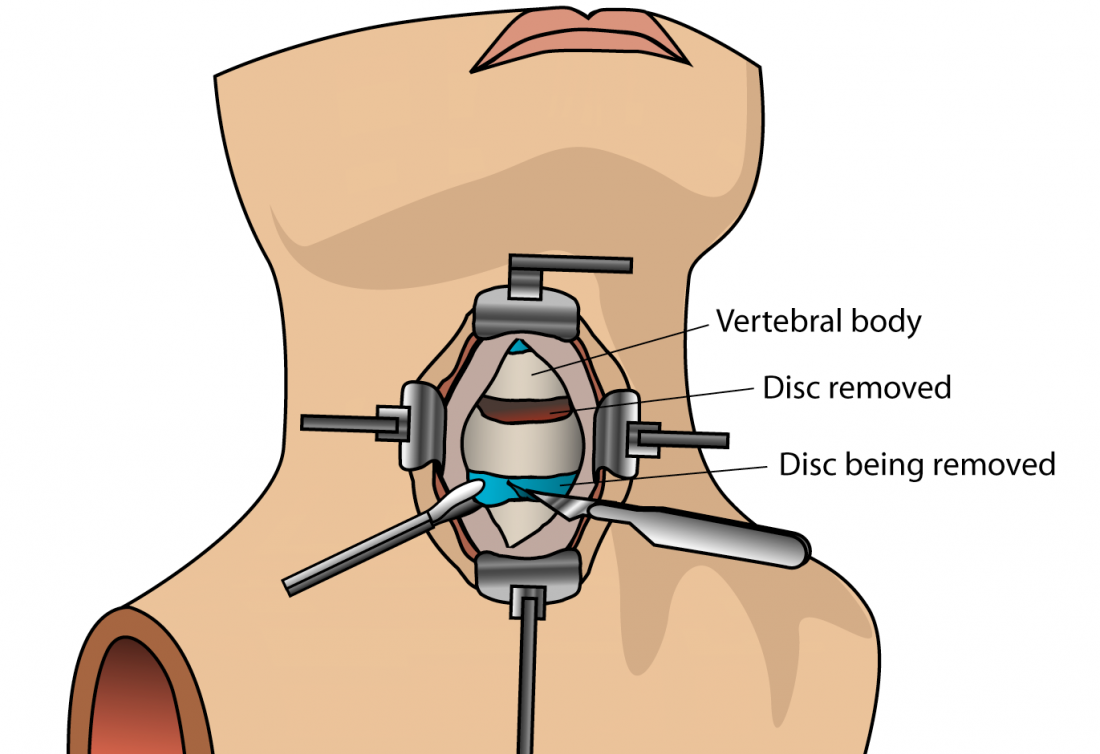
Minimally Invasive Surgery for Cervical Nerve Compression (Pinched Nerve in the Neck)
What Is This Surgery?
Microscopic Cervical Foraminotomy is a minimally invasive spine surgery performed to relieve pressure on a cervical nerve root that is being compressed as it exits the spinal canal.
The procedure is performed through a small incision at the back of the neck using an operating microscope and specialised microsurgical instruments. Unlike fusion surgery (ACDF), the affected cervical disc is usually preserved, and no implants are required in appropriately selected patients.
The goals of the surgery are to:
Relieve pressure on the compressed cervical nerve root
Reduce arm pain (cervical radiculopathy)
Improve numbness and tingling
Restore muscle strength
Preserve normal neck movement
Avoid spinal fusion whenever appropriate
Enable faster recovery with minimal muscle damage
Microscopic Cervical Foraminotomy is considered a motion-preserving minimally invasive cervical spine procedure and is an excellent option for selected patients with unilateral cervical nerve compression.
Why Is This Surgery Performed?
This surgery may be recommended for:
Cervical Foraminal Stenosis
Cervical Radiculopathy
Lateral Cervical Disc Herniation
Cervical Bone Spur (Osteophyte) causing nerve compression
This procedure is generally recommended for patients with nerve root compression causing arm symptoms, rather than patients with significant spinal cord compression (myelopathy).
Who Is A Suitable Candidate?
You may be a suitable candidate if:
✓ Symptoms persist despite medications and physiotherapy.
✓ Arm pain is more severe than neck pain.
✓ MRI confirms compression of a cervical nerve root within the neural foramen.
✓ Symptoms correlate with MRI findings.
✓ Progressive weakness develops.
✓ Conservative treatment has failed.
✓ Quality of life is significantly affected.
✓ Cervical spine stability is maintained and fusion is not required.
How Is The Surgery Performed?
Step 1
The procedure is performed under general anesthesia. The patient is positioned face down or in a sitting position depending on the surgeon's preference and surgical setup.
Step 2
A small skin incision, usually 2–3 cm, is made over the affected level at the back of the neck.
Step 3
Using specialised tubular retractors or microsurgical muscle-splitting techniques, the neck muscles are gently separated rather than cut. An operating microscope provides excellent magnification and illumination throughout the procedure.
Step 4
A small portion of bone and ligament is carefully removed to enlarge the neural foramen (the passage through which the nerve exits). Any herniated disc fragments or bone spurs compressing the nerve are removed while preserving the stability of the cervical spine.
Step 5
After confirming complete nerve decompression, the wound is closed using cosmetic sutures.
Most Microscopic Cervical Foraminotomy procedures are completed within 60–90 minutes, depending on the complexity and number of levels treated.
What Are The Advantages?
Potential benefits include:
Small cosmetic incision
Minimal muscle damage
Motion-preserving surgery (no fusion required in suitable patients)
No implants required in most cases
Reduced blood loss
Less postoperative pain
Preservation of normal neck movement
Faster recovery
Shorter hospital stay
Early mobilisation
Rapid relief of arm pain
Quicker return to daily activities
Improved quality of life
Compared with ACDF, Microscopic Cervical Foraminotomy preserves the natural movement of the operated spinal segment and avoids fusion in carefully selected patients.
What Are The Risks?
Every surgical procedure carries certain theoretical risks and potential complications. However, every effort is made to minimise these risks through meticulous surgical planning, advanced technology and comprehensive patient optimisation.
At Bombay Spine Clinic™, Microscopic Cervical Foraminotomy is performed using a high-definition operating microscope, specialised microsurgical instruments and minimally invasive muscle-preserving techniques. These technologies allow precise decompression of the affected cervical nerve while minimising disruption to surrounding muscles and normal spinal structures.
In addition, every patient undergoes a detailed pre-operative assessment. Depending on individual health requirements, this may involve evaluation and optimisation by physicians, cardiologists, anaesthetists and other specialists. This multidisciplinary approach helps improve surgical safety, recovery and overall outcomes.
Potential risks that are discussed with patients include:
Infection
Bleeding
Dural tear (CSF leak)
Nerve injury
Persistent numbness
Persistent weakness
Recurrent nerve compression
Incomplete symptom relief
Neck pain
Rare spinal instability requiring future fusion
Rare need for revision surgery
Fortunately, significant complications are uncommon when surgery is performed for appropriate indications using modern minimally invasive microsurgical techniques.
Preparing For Surgery
Before surgery:
Clinical evaluation
MRI review
CT scan (if required)
Blood investigations
Medical fitness assessment
Medication review
Anaesthesia assessment
Patients should inform their doctor regarding:
Blood thinning medications
Diabetes
Heart disease
Previous neck surgeries
Allergies
Smoking history
Smoking cessation is strongly encouraged as it promotes faster healing and better surgical outcomes.
What Happens During Hospital Stay?
Day of Surgery
Hospital admission
Surgery
Recovery room observation
Pain management
Early neurological assessment
First 24 Hours
Assisted walking
Physiotherapy instructions
Wound care guidance
Neck mobility advice
Discharge
Most patients are discharged within:
24 hours
Many patients are discharged the following day depending on their recovery.
Recovery After Surgery
Week 1
Expected symptoms:
Mild neck discomfort
Improvement in arm pain
Walking encouraged
Light daily activities permitted
Weeks 2–6
Gradual increase in activity
Neck mobility exercises
Physiotherapy if advised
Follow-up consultation
6 Weeks–3 Months
Progressive strengthening exercises
Return to routine activities
Functional recovery
Long-Term Recovery
Most patients experience significant relief of arm pain, numbness and weakness while maintaining normal neck movement. Maintaining good posture, cervical strengthening exercises and ergonomic habits helps reduce future problems.
When Can I Return To Work?
Occupation
Approximate Timeline
Desk Job
2–3 weeks
Light Work
3–4 weeks
Manual Labour
6–8 weeks
Driving
2–3 weeks
Sports
8–10 weeks
Frequently Asked Questions
1. Why do I need this surgery?
Because a cervical nerve is compressed by a disc or bone spur, causing persistent arm pain, numbness or weakness despite conservative treatment.
2. Are there alternatives to surgery?
Yes. Many patients improve with medications, physiotherapy, cervical traction and activity modification. Surgery is recommended when symptoms persist or neurological weakness develops.
3. Will I have a large scar?
No. The incision is usually only 2–3 cm and heals with an excellent cosmetic result.
4. How long will I stay in hospital?
Most patients are discharged within 24 hours after surgery.
5. When can I start walking?
Walking is encouraged within a few hours after surgery under supervision.
6. When can I climb stairs?
Most patients can climb stairs safely before discharge.
7. When can I drive?
Driving is usually permitted after approximately 2–3 weeks, once neck movements are comfortable and you are no longer taking strong pain medications.
8. When can I return to work?
Most office workers return within 2–3 weeks, while physically demanding jobs may require approximately 6–8 weeks.
9. What is the success rate?
When performed for appropriate indications, Microscopic Cervical Foraminotomy has an excellent success rate in relieving arm pain caused by cervical nerve root compression. Most patients experience significant improvement in pain, numbness and arm function.
10. Can the problem recur?
Although recurrence is uncommon, further disc degeneration or bone spur formation may occur over time. Maintaining good posture, neck muscle strength and ergonomic practices can help reduce this risk.
Myth vs Fact
Myth:
Every patient with a cervical slipped disc requires fusion surgery.
Fact:
Many patients with isolated cervical nerve root compression can be successfully treated with Microscopic Cervical Foraminotomy, preserving normal neck movement without the need for spinal fusion.
Myth:
Removing bone from the spine makes the neck unstable.
Fact:
Only a small amount of bone is removed to relieve nerve compression while preserving the stability of the cervical spine in carefully selected patients.
If you have persistent neck pain, arm pain, numbness or weakness due to cervical nerve compression, or would like a second opinion regarding your MRI findings, you may schedule a consultation or upload your MRI for specialist review.
Dr. Siddharth KatkadeConsultant Spine Surgeon
Bombay Spine Clinic™
Top of Form
Bottom of Form
Need Clinical Evaluation?
Consult with Dr. Siddharth Katkade (MBBS, MS, DNB, FASSI Germany/UK) for accurate diagnosis and personalized spine treatment.