
Detailed clinical explanations with downloadable doctor's guides for each spinal condition.
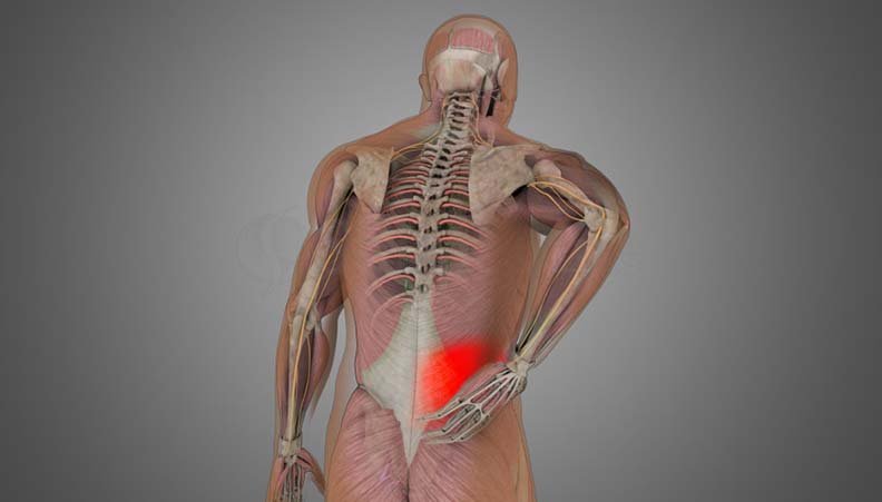
Stretching or micro-tearing of spinal ligaments and lumbar muscles caused by sudden lifting, twisting, or poor posture. Causes acute muscular spasms and localized stiffness without nerve root compression.
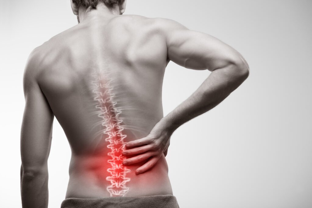
Aching discomfort aggravated by physical movement, long sitting, or standing, originating from degenerated facet joints or muscular fatigue without neurological radiation.
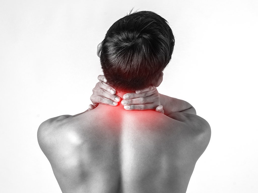
Inflammation and persistent spasm of the trapezius muscle along the upper back, neck, and shoulder blades. Often triggered by "Tech-Neck" smartphone use, prolonged laptop typing, and stress.
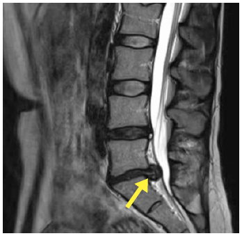
Extrusion of the soft inner gel (nucleus pulposus) through the outer annulus, directly pinching adjacent spinal nerves. Leads to acute back pain and radiating nerve pain.
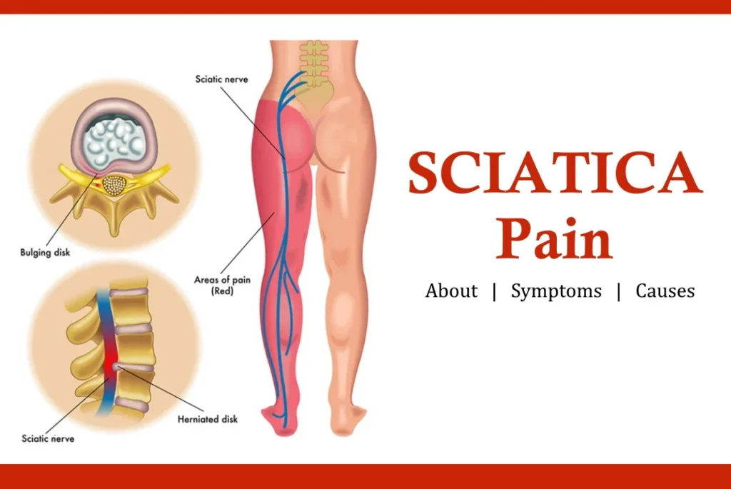
Sharp, shooting electrical pain travelling along the sciatic nerve pathway from the lower back through the buttock and down the back of the thigh to the foot and toes.
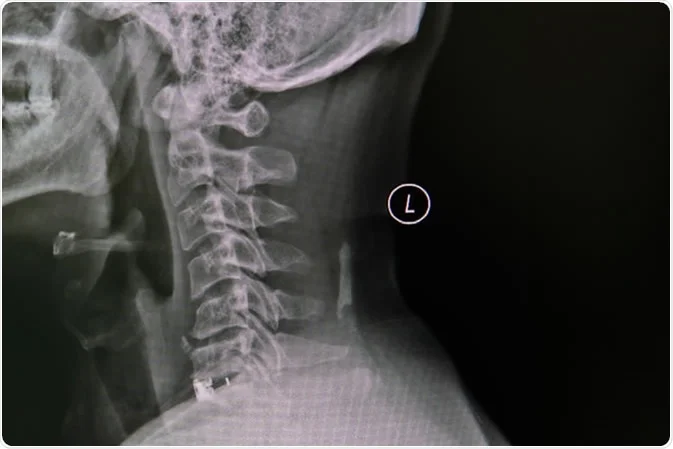
Age-related degenerative wear and tear affecting cervical vertebral discs and facet joints, leading to neck stiffness, grinding sensations, headaches, and radiating shoulder pain.
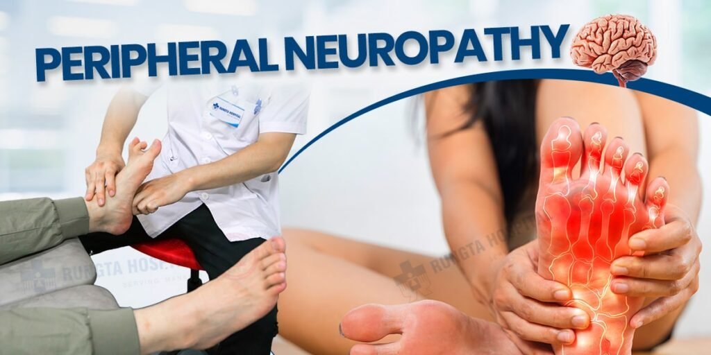
Damage to peripheral nerves (commonly associated with diabetes or vitamin B12 deficiency), causing symmetrical stocking-and-glove numbness, tingling, and hypersensitivity in feet.
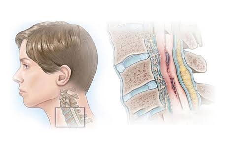
Compression of the spinal cord in the neck causing loss of fine motor hand skills (difficulty buttoning shirts, handwriting deterioration) and unsteady, drunken-like walking balance.
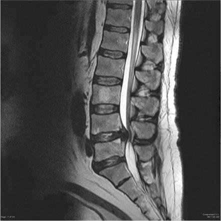
Narrowing of the spinal canal due to thickened ligaments and arthritic bone spurs, leading to neurogenic claudication—heaviness, cramping, and numbness in legs after walking a short distance.
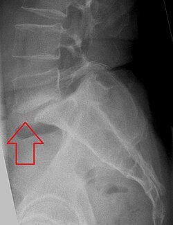
Slippage of one vertebra over the one below it (commonly L4 over L5 or L5 over S1), resulting from pars interarticularis defects (isthmic) or degenerative facet arthritis.
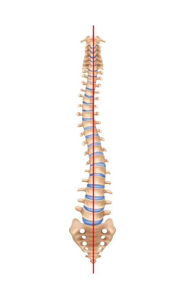
Abnormal lateral sideways curvature of the spine (forming an 'S' or 'C' curve). Can be adolescent idiopathic or adult degenerative scoliosis with uneven shoulders or hips.
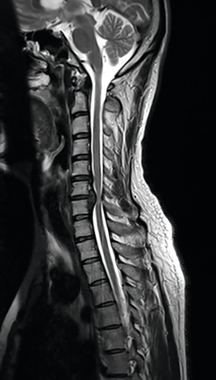
Excessive outward curvature of the thoracic spine leading to hunching or slouching posture. Caused by Scheuermann's disease, osteoporotic collapse, or postural muscle weakness.
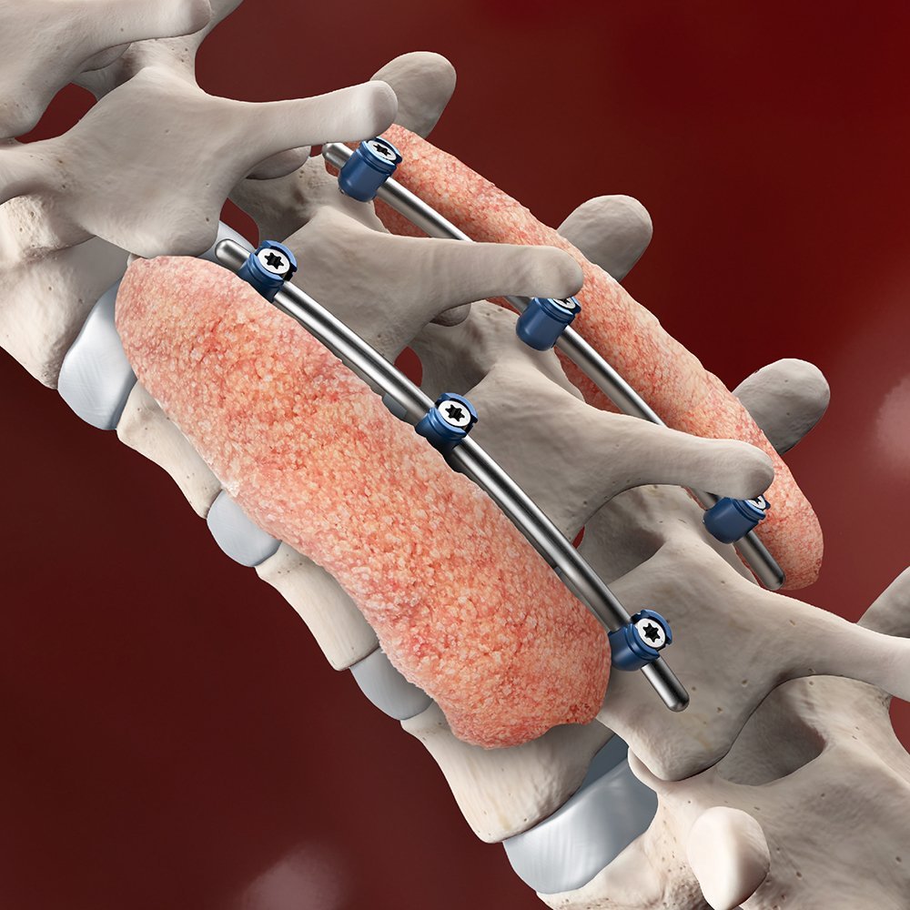
Progressive loss of bone mineral density causing porous, brittle vertebrae vulnerable to sudden compression fractures even from mild coughing or trivial falls.
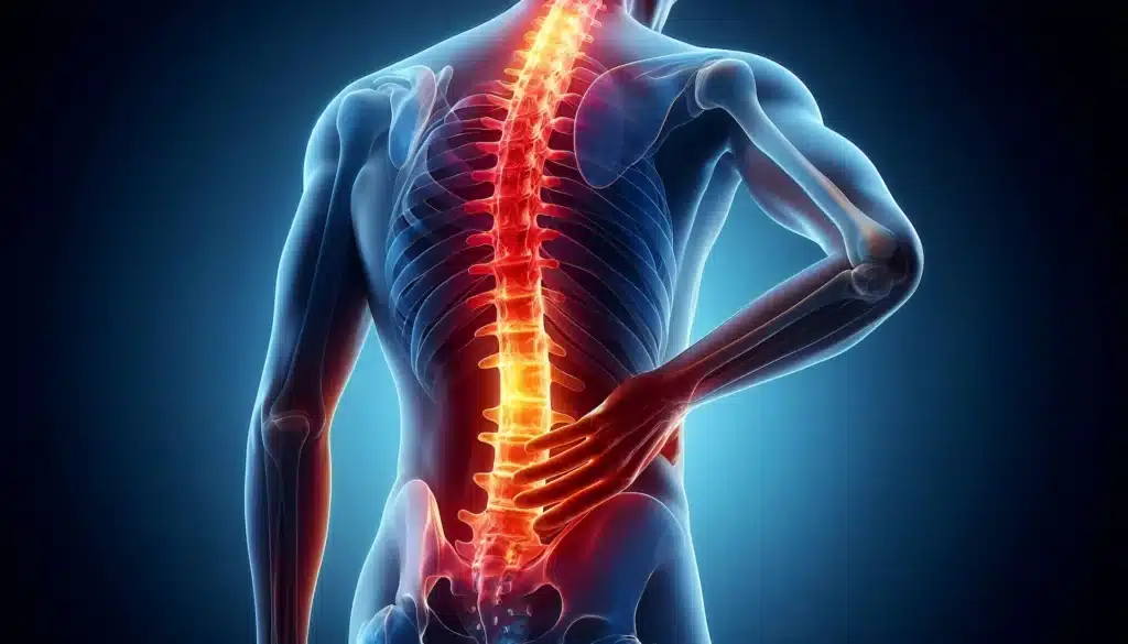
Vertebral wedge compression, burst, or flexion-distraction fractures resulting from vehicular accidents, falls from heights, or severe osteoporosis.

Bacterial infection of vertebral bodies and intervertebral disc spaces (spondylodiscitis / epidural abscess), causing relentless night pain, fever, and elevated ESR/CRP.
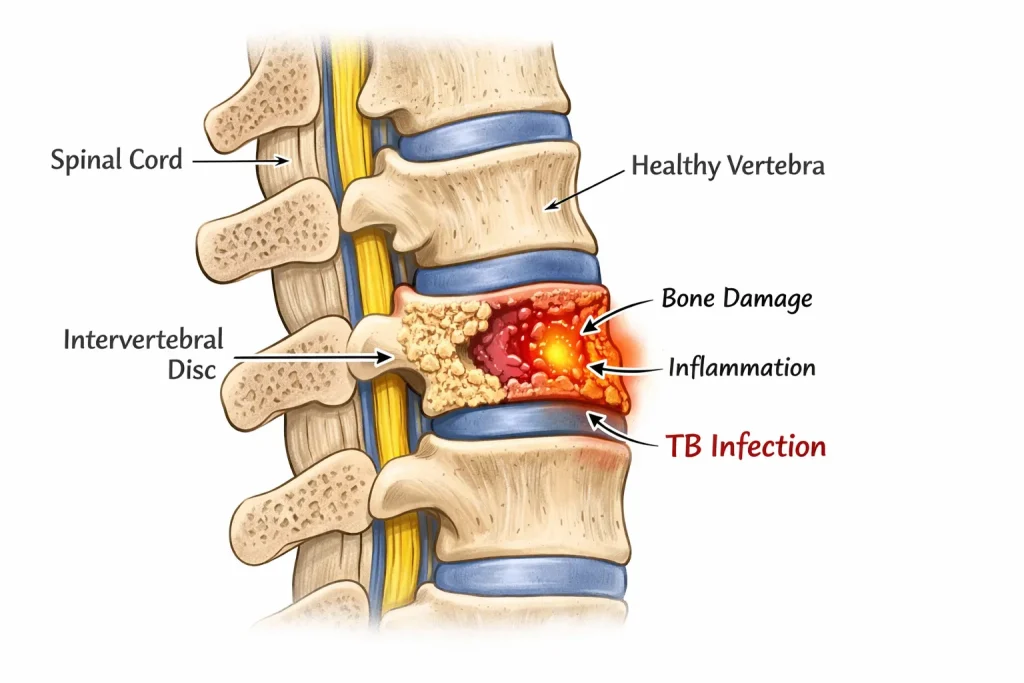
Mycobacterium tuberculosis infection attacking spinal vertebrae and intervertebral discs, leading to cold abscesses, severe kyphotic collapse, and risk of paraplegia.
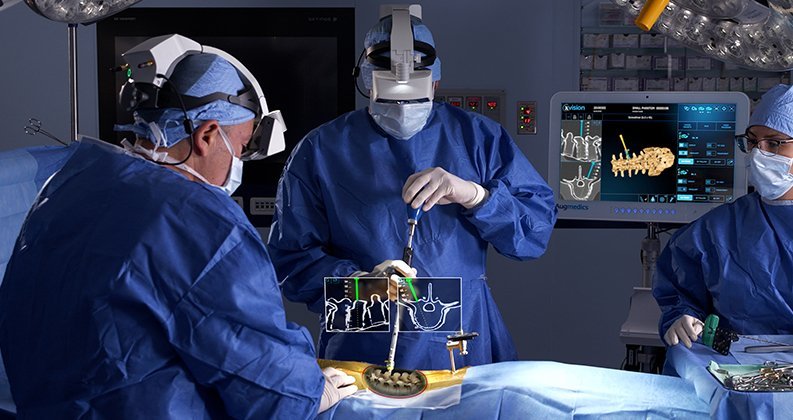
Primary spinal bone neoplasms (e.g. osteoblastoma, hemangioma, schwannoma) or metastatic lesions. Characterized by severe unremitting rest pain and progressive neurological deficits.
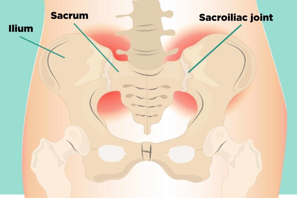
Inflammation of one or both sacroiliac joints connecting the lower spine to the pelvis. Causes sharp buttock pain, groin discomfort, and morning stiffness mimicking sciatica.
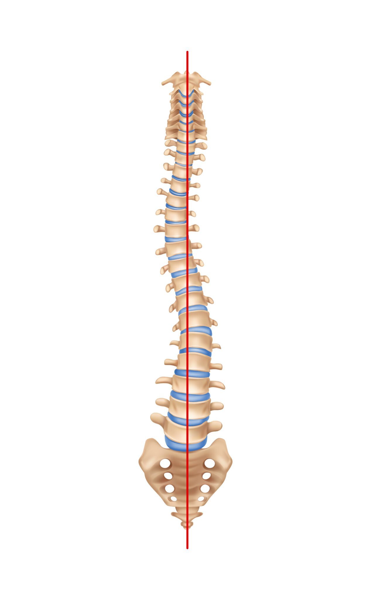
Chronic inflammatory arthritis linked to HLA-B27, causing progressive fusion of spinal vertebrae ("Bamboo Spine") and severe early morning stiffness that improves with activity.
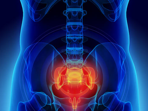
Localized pain at the tailbone (coccyx), exacerbated significantly when sitting on hard surfaces, leaning backwards, or rising from a chair. Commonly follows a fall on the buttocks.
While most back pain is non-emergency, certain neurological symptoms indicate severe spinal cord or nerve root compromise and require immediate emergency medical evaluation:
Sudden loss of bowel or bladder control (incontinence or urinary retention) accompanied by numbness in the saddle/groin area. Requires emergency decompression within 24 hours.
Sudden weakness where you cannot lift the front of your foot, causing tripping or dragging your toes while walking.
Severe deep bone pain that does not ease with rest or changing positions, often waking you from sleep (suspicion of infection or tumor).
Dropping objects, difficulty buttoning clothes, or staggering while walking (signs of Cervical Myelopathy).
Comprehensive home physiotherapy and post-surgical rehabilitation regimens prescribed by Dr. Siddharth Katkade.
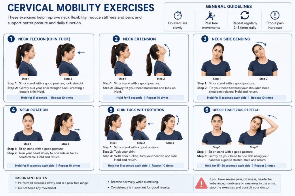
Gentle flexion, extension, lateral side bend, and slow rotations (10 reps x 2 sets daily) to improve range of motion in stiff cervical facet joints and relieve tension headaches.
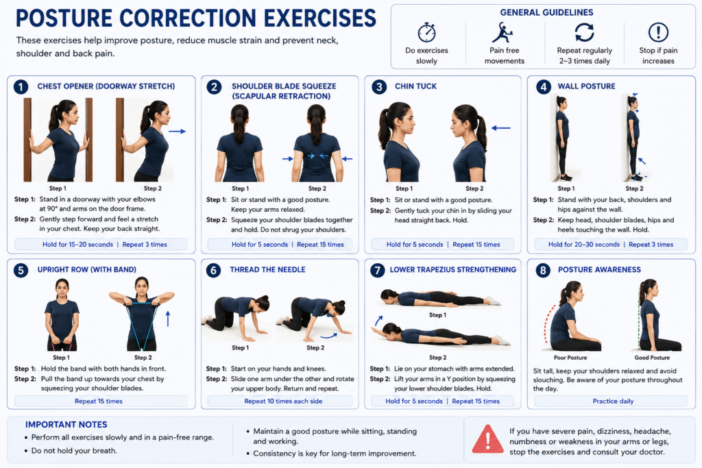
Chin tucks, scapular retractions, and wall angels to reverse rounded shoulders and Tech-Neck forward head posture, re-aligning your center of gravity.
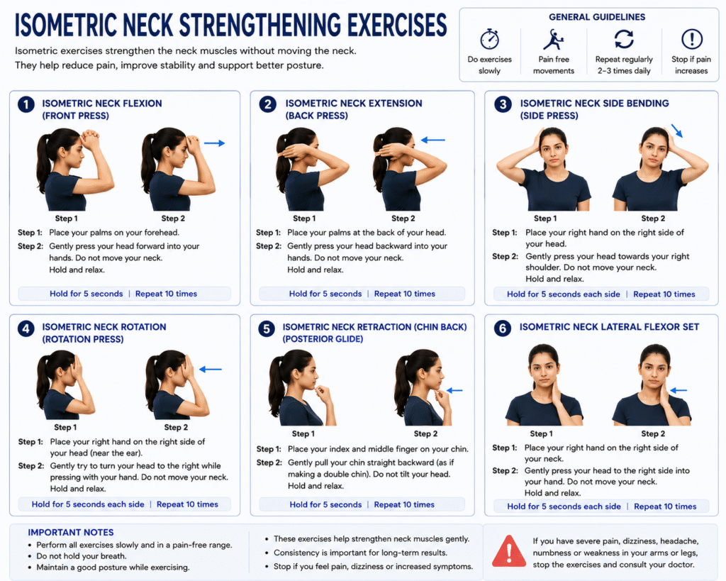
Pushing head into palm (forehead, back, right, left) for 5-10 second holds without moving the neck. Builds deep cervical flexors without irritating disc spaces.
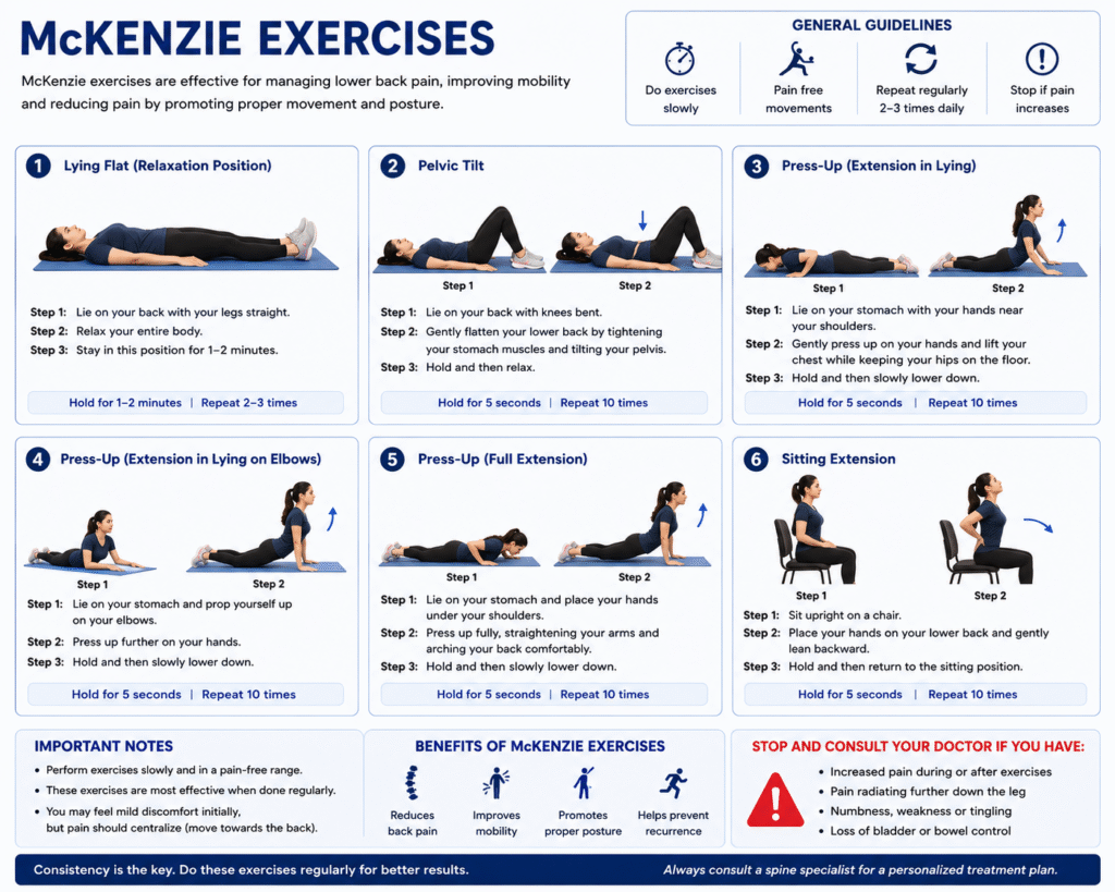
Prone on elbows and prone press-ups designed to centralize posterior disc bulges, drawing sciatica pain back out of the leg into the central lower back.
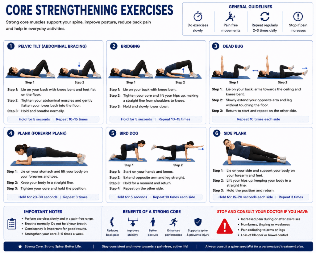
Pelvic tilts, supine bridging, bird-dog, and abdominal draw-ins that strengthen transverse abdominis and lumbar multifidus muscles to unload the lumbar discs.
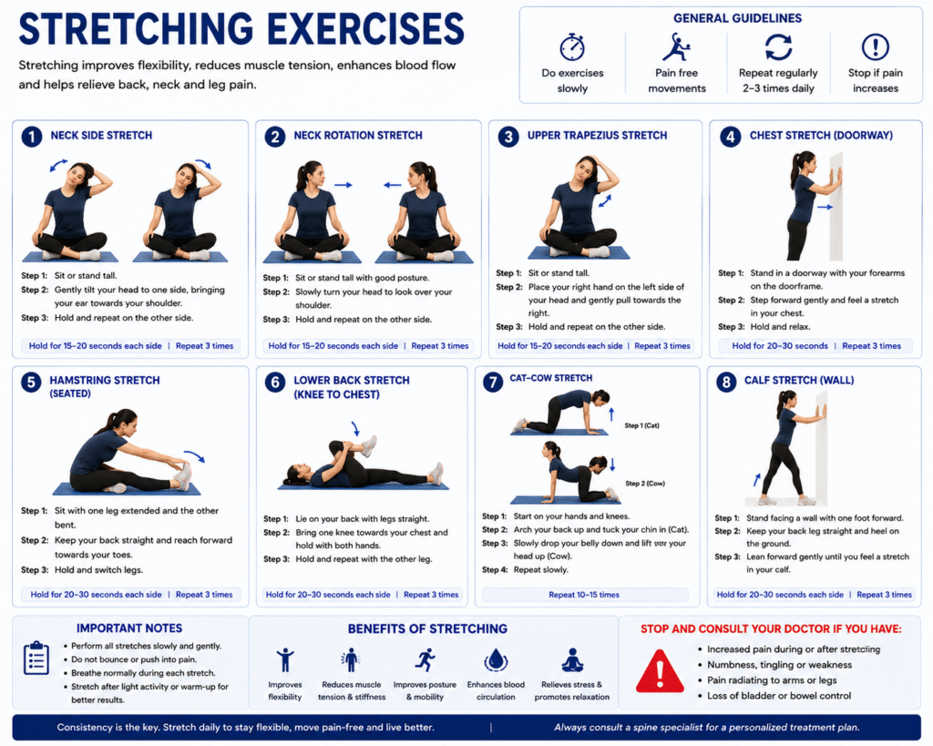
Single knee-to-chest, double knee-to-chest, cat-camel mobility stretch, and hip flexor stretches to reduce pelvic tilt and muscle tightness.
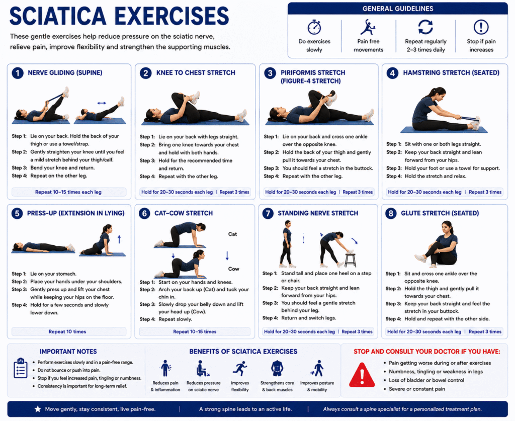
Figure-4 piriformis stretch, hamstring nerve glides, and seated neural mobilization to reduce nerve tethering and improve neural vascularity.
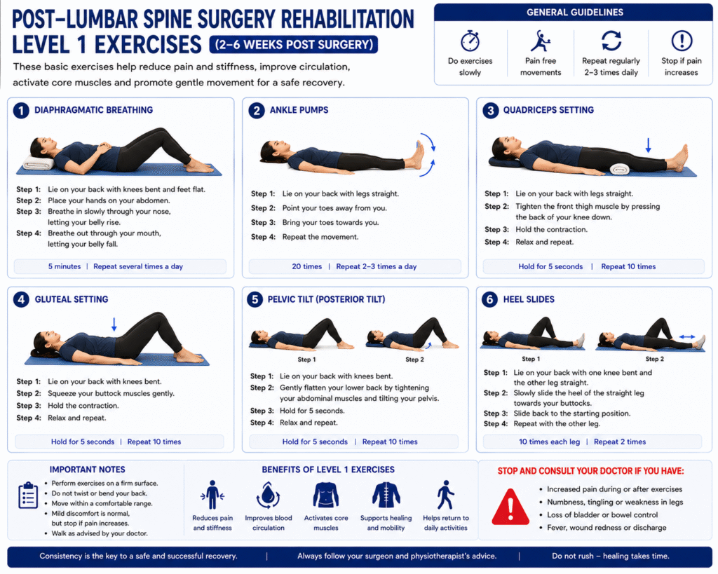
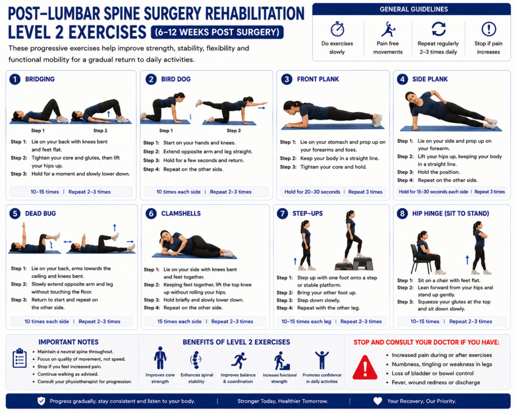
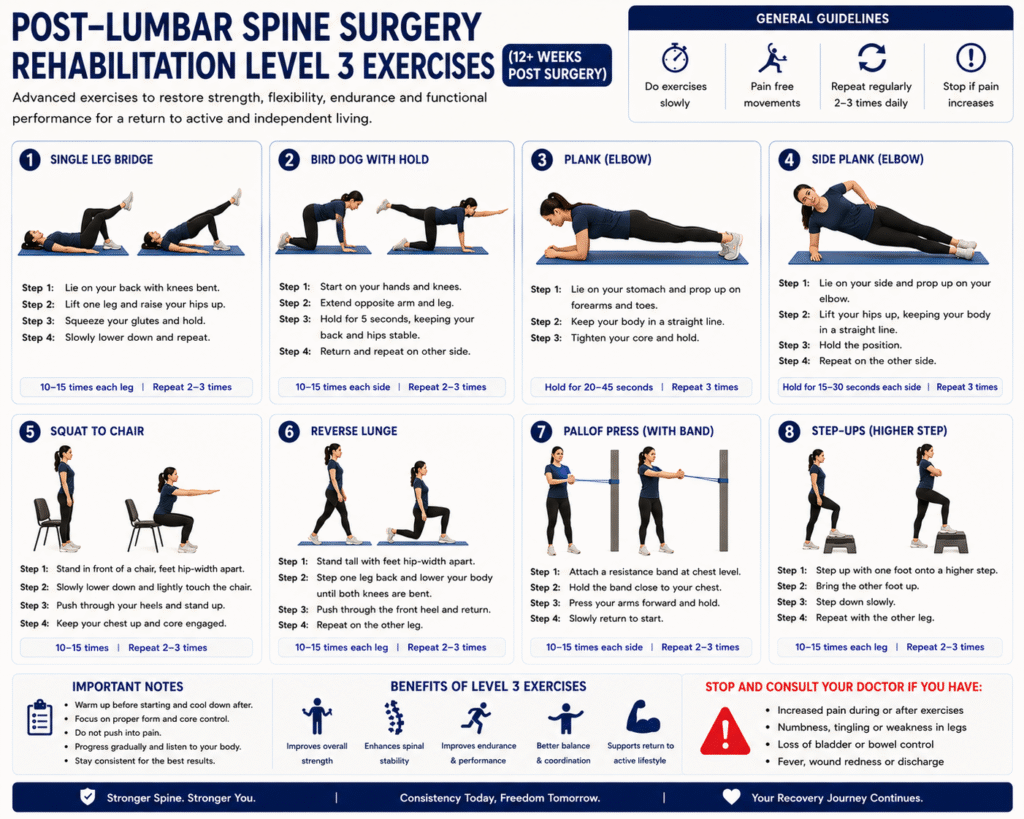
Phased rehabilitation guiding patients step-by-step from safe mobilization and log-rolling in week 1 to advanced core endurance and active sport return by month 3.
A bulge is a generalized symmetric swelling of the disc beyond the bone margin (often normal aging). A herniation/extrusion is an actual focal rupture where the gel escapes and physically pinches the spinal nerve root.
The thecal sac protects the spinal cord and nerve bundle. When discs or facet bones indent the sac or narrow the neural foramen (exit gate for leg nerves), it produces radiating sciatica pain.
Modic changes represent bone marrow edema / inflammation adjacent to degenerated discs. Osteophytes are smooth bone spurs formed by the body attempting to stabilize an unstable spinal segment.
"Treat the patient, not just the MRI scan." Many healthy individuals have painless MRI disc bulges. Dr. Siddharth Katkade correlates your exact symptoms with the imaging to ensure no unnecessary surgeries are ever performed.
A stepped care approach: exhaust conservative options first before considering precision keyhole surgery.
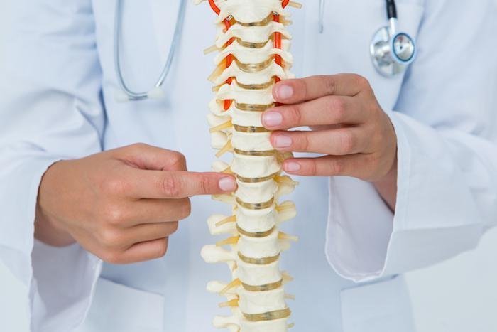
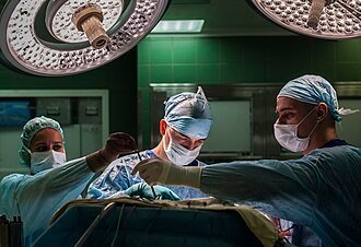
State-of-the-art operative interventions performed by Dr. Siddharth Katkade.
Routine blood tests (CBC, PT/INR, kidney/liver profiles), ECG, chest X-ray, and pre-anesthetic check-up (PAC) by the anesthesia team.
Inform your surgeon about blood thinners (Aspirin, Clopidogrel, Warfarin), which are typically paused 5-7 days prior under cardiologist guidance.
Strict fasting (no food or water) for 6 to 8 hours prior to the scheduled surgery time to ensure safe anesthesia induction.
Keep dressing dry and clean. Waterproof dressings allow showering. Stitches or Band-Aid strips are evaluated at your 10-12 day follow-up.
Follow the "BLT" rule for 4-6 weeks: No Bending forward, Lifting >3kg, or Twisting the spine.
Walking is the best medicine after spine surgery. Walk 10-15 minutes 3 to 4 times daily on flat ground, gradually increasing duration.
Car travel as a passenger is safe for short trips. For flights or long train journeys, wait 2-3 weeks, break up sitting every hour, and avoid lifting heavy luggage.
Work from home / desk jobs can typically be resumed within 7 to 14 days. Heavy physical labor or strenuous jobs require 6 to 12 weeks of structured rehab.
Clinic visits scheduled at Day 12 (wound check), Week 6 (physiotherapy progression), and Month 3 (full clearance & X-ray review).
Dairy, ragi (finger millet), paneer, tofu, sesame seeds, almonds, and leafy greens for strong vertebrae.
Morning sunlight exposure (20 mins), fortified foods, egg yolks, mushrooms, and prescribed supplements.
Eggs, lentils, sprouts, chicken, fish, and paneer to rebuild surgical tissue and repair muscles.
Drink 2.5–3 liters of water daily. Intervertebral discs rely on water diffusion for shock absorption.
Answers categorized across key patient inquiries.
Download official, publication-ready PDF guides with clinic letterhead for all conditions, surgical procedures, checklists, and diet charts:
Educational video library featuring Dr. Siddharth Katkade explaining spine surgical techniques, 3D animations, exercise demonstrations, and patient recovery walkthroughs.
We are here to assist you at every step of your spine journey.
Have questions about your symptoms or need a reliable second opinion on your MRI?